Leg Anatomy Muscles Ligaments And Tendons - ANATOMY - Leg anatomy muscles ligaments and tendons :
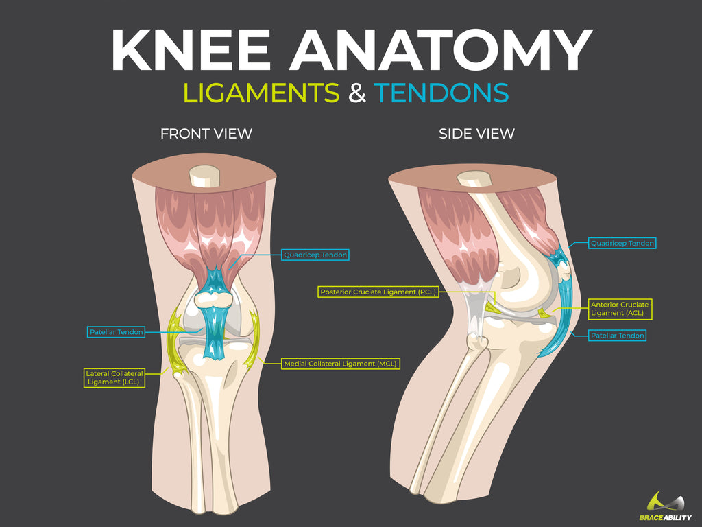
Leg Anatomy Muscles Ligaments And Tendons - ANATOMY - Leg anatomy muscles ligaments and tendons :. The term has also been used in reference to areas of thickened peritoneal folds that are important in anchoring adjacent viscera to each other as well as the abdominal wall. One of the most important tendons is the quadriceps tendon. The tarsal bones are found near the. This lies on the front of the knee and connects the quadriceps muscles of the thigh to the tibia via the patella and patellar ligament (or tendon). The calf muscles, through the achilles tendon, are the main plantarflexors of the ankle which pulls the foot down.
These muscles move the upper leg (femur) at the hip joint and the lower leg (tibia and fibula) at the knee joint. Tendons connect muscles to bones, while ligaments connect bones to other bones. Learn about the muscles, tendons, bones, and ligaments that comprise the knee joint anatomy. It is made up of bones, muscles, tendons, ligaments and 100 other which are designed o allow the foot to balance the body on two legs. A joint capsule is a watertight sac that surrounds a joint.
Ankle Anatomy - Be In Motion Physiotherapy from www.beinmotion.ca Leg anatomy muscles ligaments and tendons : Possibly the most important tendon in terms of mobility is the achilles tendon. Ligaments are structures that connect two bones together. Tendons attach muscle to bone. There are four muscles in the anterior compartment of the leg. This lies on the front of the knee and connects the quadriceps muscles of the thigh to the tibia via the patella and patellar ligament (or tendon). The calf muscles, through the achilles tendon, are the main plantarflexors of the ankle which pulls the foot down. Tendons attach muscle to bone.
These are called the cruciate ligaments and consist of the anterior cruciate ligament and the posterior cruciate ligament.
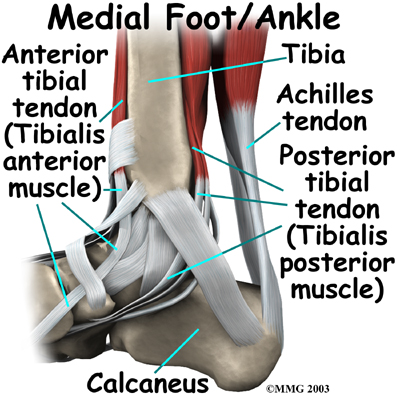
Ligaments also support the lower end of the leg where it forms a hinge for the ankle. To ensure your body moves smoothly with a minimum of friction, muscles are enveloped in a slippery skin like tissue called fascia. This important tendon in the back of the calf and ankle connects the plantaris, gastrocnemius, and soleus muscles to. The calf muscles, through the achilles tendon, are the main plantarflexors of the ankle which pulls the foot down. A joint capsule is a watertight sac that surrounds a joint. Tendons vary in size and are somewhat elastic and attach bones to muscles. Both cross the ankle, but the peroneus longus wraps underneath the cuboid crossing the plantar aspect of the foot as well, and inserts at the base of the first metatarsal. Tendons attach muscle to bone. 12 photos of the muscles and tendons of the leg. Learn about the muscles, tendons, bones, and ligaments that comprise the knee joint anatomy. There are over two dozen gorgeous and painstakingly detailed illustrations on this chart, from the extensor hallucis longus to the flexor digitorum brevis. Ligaments are soft tissue structures that connect bones to bones. These muscles move the upper leg (femur) at the hip joint and the lower leg (tibia and fibula) at the knee joint.
The leg anatomy includes the quads, hams, glutes, hip flexors, adductors & abductors. Ligaments also support the lower end of the leg where it forms a hinge for the ankle. 4.3.1 similar to what is observed at the wrist, tendons at the ankle region passing from the leg into the in this manner, the two muscles form a tendinous sling under the foot, which serves to support. Leg anatomy muscles ligaments and tendons : This chart is perfect for educating medical students or for…
MediVisuals Normal Foot Anatomy Exhibits from medivisuals1.com The leg anatomy includes the quads, hams, glutes, hip flexors, adductors & abductors. It is made up of bones, muscles, tendons, ligaments and 100 other which are designed o allow the foot to balance the body on two legs. The term has also been used in reference to areas of thickened peritoneal folds that are important in anchoring adjacent viscera to each other as well as the abdominal wall. There are over two dozen gorgeous and painstakingly detailed illustrations on this chart, from the extensor hallucis longus to the flexor digitorum brevis. Originating below and beneath the gastrocnemius is the soleus muscle, which extends your foot when your knee is bent. Leg anatomy muscles ligaments and tendons : The tendons of the edl can be palpated on the dorsal surface of the foot. You hear them referred to as your gams, poles or limbs. but, whatever you call them, your legs are composed of bones, muscles, tendons and ligaments.
Both the acl and the pcl function to stabilize the knee from front to back.
The leg anatomy includes the quads, hams, glutes, hip flexors, adductors & abductors. The tendons of the edl can be palpated on the dorsal surface of the foot. Ligaments connect two or more bones together and help stabilize joints. The tarsal bones are found near the. Muscles, either individually or in groups, are supported by fascia. A joint capsule is a watertight sac that surrounds a joint. Related posts of muscle, tendons and ligaments of leg human. Leg anatomy muscles ligaments and tendons : Tendons vary in size and are somewhat elastic and attach bones to muscles. This lies on the front of the knee and connects the quadriceps muscles of the thigh to the tibia via the patella and patellar ligament (or tendon). Leg anatomy muscles ligaments and tendons. Ligaments are structures that connect two bones together. Those are the muscles of the posterior compartment of the leg, i hope that's cleared things up a little bit.
The two main calf muscles, gastrocnemius and soleus, run down the back of the calf and join together to form a strong, thick tendon, the achilles tendon, that attaches to the back of the heel. The calf muscles, through the achilles tendon, are the main plantarflexors of the ankle which pulls the foot down. Tendons connect muscles to bones, while ligaments connect bones to other bones. Both the acl and the pcl function to stabilize the knee from front to back. To better understand foot and leg muscle/tendon injuries, it is important to appreciate the basic elements that enable your body parts to move.
Pain Behind Knee | Why it Hurts in Back of or Under your ... from cdn.shopify.com The two main calf muscles, gastrocnemius and soleus, run down the back of the calf and join together to form a strong, thick tendon, the achilles tendon, that attaches to the back of the heel. Muscles, either individually or in groups, are supported by fascia. 4.3.1 similar to what is observed at the wrist, tendons at the ankle region passing from the leg into the in this manner, the two muscles form a tendinous sling under the foot, which serves to support. Tendons connect muscles to bones, while ligaments connect bones to other bones. It is made up of bones, muscles, tendons, ligaments and 100 other which are designed o allow the foot to balance the body on two legs. Muscles, tendons, and ligaments run along the surfaces of the feet, allowing the complex movements needed for motion and balance. Sacrospinous ligament (dorsal view) the latter ligament has several attachments to the posterior sacroiliac ligaments, lower transverse tubercles of the sacrum, the posterior superior iliac spine, the proximal part of the. A joint capsule is a watertight sac that surrounds a joint.
The two main calf muscles, gastrocnemius and soleus, run down the back of the calf and join together to form a strong, thick tendon, the achilles tendon, that attaches to the back of the heel.
This important tendon in the back of the calf and ankle connects the plantaris, gastrocnemius, and soleus muscles to. Tendons attach muscle to bone. Related posts of muscle, tendons and ligaments of leg human. To ensure your body moves smoothly with a minimum of friction, muscles are enveloped in a slippery skin like tissue called fascia. Leg anatomy muscles ligaments and tendons : Leg anatomy muscles ligaments and tendons : Dr donald a ozello dc of championship chiropractic in las vegas, nv is the author of running: Both the acl and the pcl function to stabilize the knee from front to back. Two of these ligaments are in the center of the joint, and they cross each other. Originating below and beneath the gastrocnemius is the soleus muscle, which extends your foot when your knee is bent. It provides the power necessary to straighten the knee. Ligaments connect two or more bones together and help stabilize joints. Related posts of muscles and tendons of the leg back muscles anatomy video.
0 Response to "Leg Anatomy Muscles Ligaments And Tendons - ANATOMY - Leg anatomy muscles ligaments and tendons :"


0 Response to "Leg Anatomy Muscles Ligaments And Tendons - ANATOMY - Leg anatomy muscles ligaments and tendons :"
Post a Comment